
The electron microscope is an essential tool for studying the microscopic world, especially bacteria. But what makes it so useful compared to traditional light microscopes when it comes to studying bacterial cells?
The extremely high magnification and resolving power of electron microscopes allow visual penetration of bacterial structures like cell walls which is not possible with light microscopy.
1. High Magnification Reveals Critical Details
With magnifications over 500,000 times, electron microscopes can reveal incredibly minute details of bacterial cells. Structures like pili and flagella, the bacterial capsule, and internal components like ribosomes become visible. This ultra-high magnification enables researchers to observe bacterial shapes, sizes, surface features, and internal organization at the nanometer scale.
Seeing this level of detail is critical for identifying different bacterial species, studying how bacterial cells function, and observing cellular changes in response to antibiotics or environmental factors. Light microscopes lack the magnification to show such small-scale structures clearly.
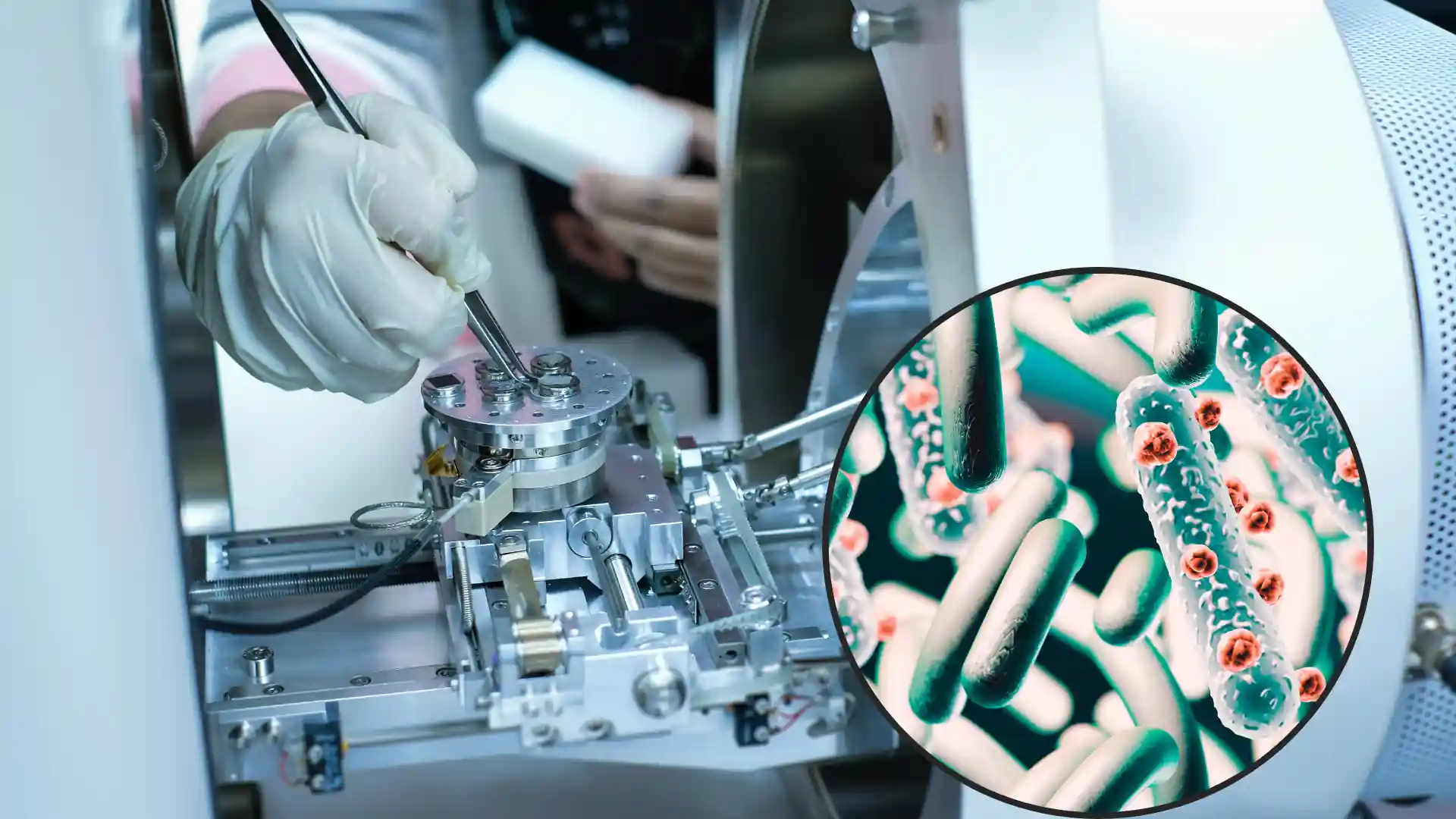
2. Extremely High Resolving Power Of EMs Can Map Hidden Structures Within Bacterial Cells
In addition to high magnification, electron microscopes have extremely high resolving power. Resolving power refers to the minimum distance between two points that can still be distinguished as separate entities.
For electron microscopes, this is around 0.2 nanometers, up to 1000 times higher than light microscopes. This enables visualization of the smallest bacterial components like surface proteins and even individual atoms.
With this resolving power, electron microscopy can reveal bacterial cell structures and features impossible to discern under light microscopy. This includes complex surface layers like capsules, fimbriae, and S-layers, diverse bacterial appendages, and intricate internal organization.
3. Imaging Ability of Ems Help Study the Dynamic Processes and Interactions Within Cells
The imaging capabilities of electron microscopy also enable researchers to observe dynamic bacterial processes including cell division, secretion of proteins or toxins, biofilm formation, and interactions with antimicrobials or host cells.
Capturing events like phagocytosis, bacterial conjugation, and viral infections would be impossible without the direct visualization offered by electron microscopy.
Frequently Asked Questions
What Sample Preparation Is Needed for Electron Microscopy of Bacteria?
Bacteria samples generally require chemical fixation, dehydration, and staining with heavy metal solutions to provide enough contrast for imaging. Samples also must be very thin, often achieved through sectioning or sputter coating.
What Are Some Limitations of Electron Microscopy for Bacterial Research?
The extensive sample preparation can artefactually alter cell morphology. Imaging only provides static snapshots. And electron beams can damage delicate samples. Fluorescence microscopy can complement EM by showing dynamic processes in living cells.
Can Individual Molecules Be Resolved with Electron Microscopy?
In some cases, yes. Cryo-electron microscopy involves flash-freezing samples to image biomolecules like proteins in their native state. Recent advances now enable atomic-level resolution of individual proteins and macromolecular complexes.
How Does Immunoelectron Microscopy Help Identify Proteins in Bacterial Cells?
Antibodies with attached electron-dense particles can bind to specific target proteins in the cell. Their location is then detectable by electron microscopy, allowing localization of proteins within bacterial structures.