
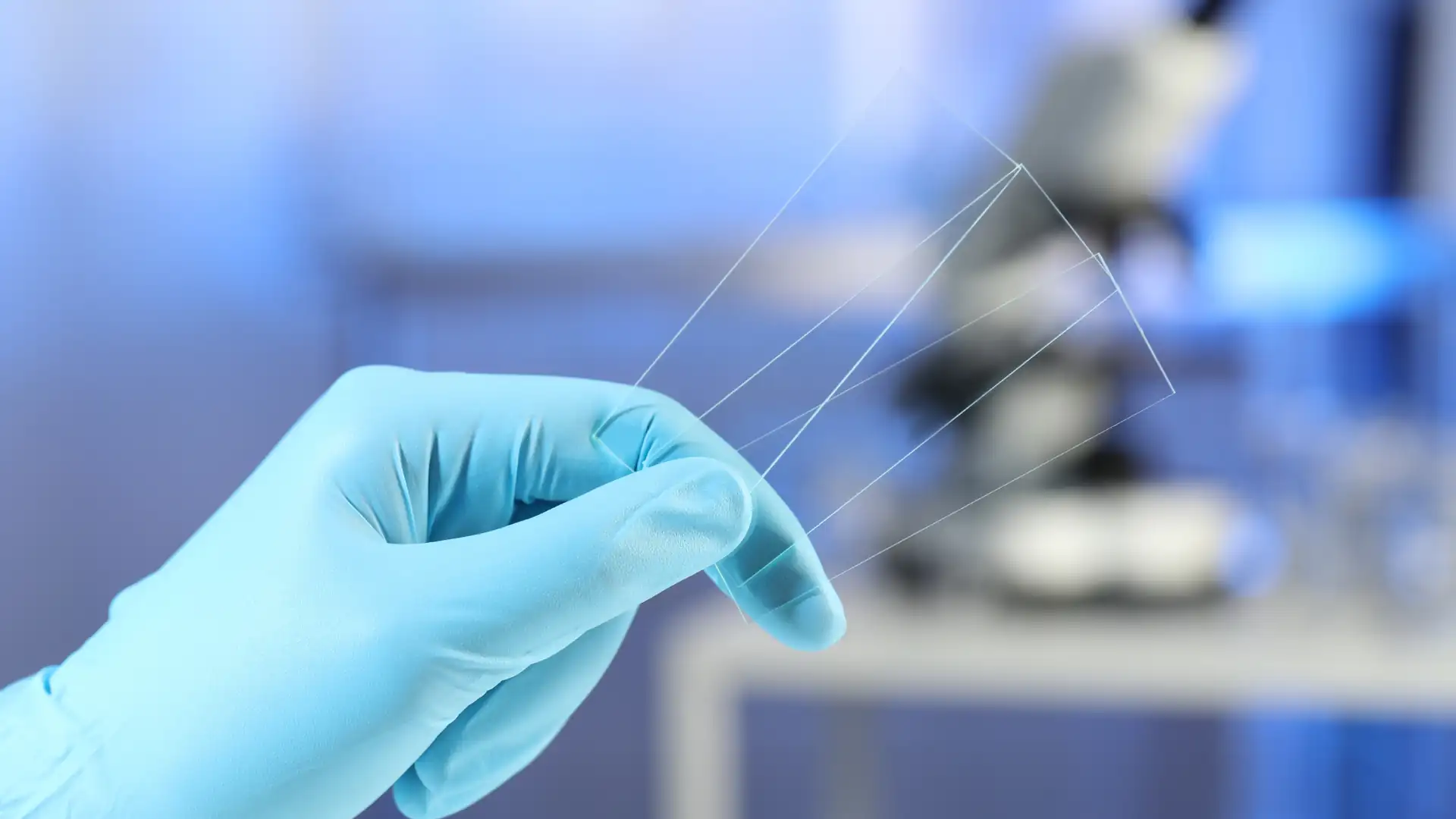
Have you ever wondered what all those tiny cells and microorganisms look like under a microscope? Preparing your microscope slides is an easy and fun way to explore the microscopic world. In this beginner’s guide, we’ll walk you through the steps for preparing three major types of microscope slides: wet mounts, dry mounts, and heat-fixed slides.
The key to getting a good look at your samples under the microscope is properly preparing the slides. With some basic materials and following a few simple techniques, you’ll be exploring the captivating microworld in no time!
1. Wet Mount Slides
Wet mount slides, also known as temporary mounts, are easy to prepare with fresh specimens like plant sections or microorganisms. Here’s what you need and how to make them:
Materials Required
- Microscope slides and coverslips
- Dropper pipettes
- Distilled water/saline solution
- Tweezers
- Specimens (onion skin, leaf sections, protozoa culture, etc.)
Step-by-Step Procedure For Wet Mount Slide Preparation
- Place a small drop of water or saline in the center of a clean microscope slide using a dropper pipette. The drop should be slightly smaller than the size of the coverslip.
- Using tweezers, carefully transfer your specimen (a thin onion skin or leaf section works well) onto the drop of liquid. Make sure the sample is completely immersed in the fluid.
- Gently lower a coverslip at an angle onto the sample to avoid trapping air bubbles. Apply slight pressure on the coverslip once it contacts the liquid to further eliminate any air pockets.
- Carefully blot away any excess liquid from under the coverslip using a paper towel or tissue paper. Your wet mount slide is now ready!
- Place the prepared slide on the microscope stage and secure it with stage clips. Start observing under a power objective lens first before switching to a higher magnification lens.
Common Wet Mount Samples
Onion skin, leaf sections, pond water samples, and protozoa cultures like paramecium, euglena, amoeba, etc. are commonly observed as wet mounts. The aqueous environment keeps the samples alive for examination over a short period before they eventually dry out.
2. Dry Mount Slides
Dry mount slides are suitable for examining specimens like insect parts, hairs, and fibers that don’t require an aqueous medium. Here are the materials and methods:
Materials Required
- Microscope slides and coverslips
- Mounting medium (clear nail polish or commercial slide mounting fluid)
- Specimens (insect legs, pet hairs, plant fibers, etc.)
- toothpicks
Step-by-Step Procedure For Dry Mount Slide Preparation
- Place your thoroughly clean and dry specimen (a single hair or insect leg works well) close to the center of a microscope slide using a toothpick or tweezers.
- Add a tiny drop of mounting medium like clear nail polish or a commercial slide mounting fluid on top of the specimen.
- Slowly lower a coverslip onto the drop, starting from one edge and lowering it gently to prevent air bubbles.
- Use the toothpick to scrape away any excess mounting fluid around the edges of the coverslip.
- Allow the mountant to thoroughly dry and harden overnight before observing under the microscope. Store the slide flat or vertically in a slide box.
Common Dry Mount Samples
Insect body parts, pet hair, plant fibers, crystals, sand grains, and skin cells like cheek cells scraped from the inner lining of the mouth are commonly prepared as permanent dry mounts.
3. Heat Fixed Slides
Heat fixing kills and adheres specimens like single-cell microorganisms onto slides before staining procedures. Here are the required materials and protocol:
Materials Required
- Microscope slides
- Heat fixative solutions – 95% ethyl alcohol/methanol
- Stains (crystal violet, safranin, etc.)
- Bacteriological loop/ Culture from broth
- Bunsen burner
- Distilled water
- Coverslips
Step-by-Step Procedure For Wet Heat Fixed Slide Preparation
- Prepare a heat-fixative solution of 95% ethyl alcohol or methanol. Dip the slide into the solution briefly and ignite the excess solution on the slide by passing over a Bunsen burner flame.
- Allow the heated slide to cool for a few seconds before aseptically smearing or spreading the microbial sample onto the slide using an inoculation loop.
- Pass the smeared slide over the Bunsen flame 2-3 times to heat-fix the cells onto the slide’s surface. Do not overheat.
- Flood the heat-fixed smear with a suitable staining solution like crystal violet or safranin for 30-60 seconds. Rinse gently with distilled water.
- Blot dry and observe under an oil immersion objective lens after adding a drop of immersion oil and securing with a coverslip.
Common Heat Fixed Samples
Bacteria, yeasts, protozoa, and tissue culture cells are commonly heat-fixed on slides before Gram staining, endospore staining, acid-fast staining, etc. This burst fixes the microbes onto the slides for further processing.
The key steps for preparing a good microscopic slide are: mounting the specimen properly on the slide’s center without any folds or air bubbles using a suitable mounting medium based on the sample type (wet/dry/heat fixed), evenly spreading out the sample, fixing onto the slide if required through heat by passing over a Bunsen burner flame, then applying suitable stains for enhancing contrast and finally observing under the microscope under optimal magnification after placing the slide on the stage and securing with stage clips.
Conclusion
Slide preparation is an integral part of operating a microscope. Learning to successfully prepare the microscope slides and exploring the minuscule world can give you a massive sense of accomplishment and a boost of confidence especially after you master techniques like wet mounting, dry mounting, and heat fixing according to each specimen’s properties.
So, what’s the delay for? Grab your microscope and all the needed materials mentioned above and get started with slide preparation.