Have you ever wondered how scientists can study cells, bacteria, or even atoms? The answer lies in the microscopic world unlocked by special instruments called microscopes. These powerful devices use lenses, beams of electrons, or precise scanning tips to magnify tiny objects up to a million times their actual size, allowing researchers to probe deeper into their structure, function, and secrets. But with so many types available today, which are the most essential for advancing scientific understanding? I examined the field to spotlight the 5 game-changing microscopes you should know about.
We rely on microscopes far more than you’d expect, from developing life-saving medications, ensuring food safety, diagnosing diseases, investigating forensic evidence, and pushing the frontiers of materials science. As innovations continue apace, these remain the trailblazing technologies no lab can live without!
1. Light Microscopes
We kick off our microscopic journey with light microscopes – the first type invented around 1590 and still ubiquitous today. Earlier versions simply used glass lenses and the visible light spectrum to magnify tiny structures. But could you guess the magnification limit for basic optical microscopes? It’s estimated to be 1500x, constrained by the wavelengths of light itself.
Later technological leaps incorporated more advanced illumination, optics, digital sensors, and software to enhance the imaging capabilities of these devices. Nonetheless, for routine examination of cell and tissue samples, 3 main types continue their enduring reign:
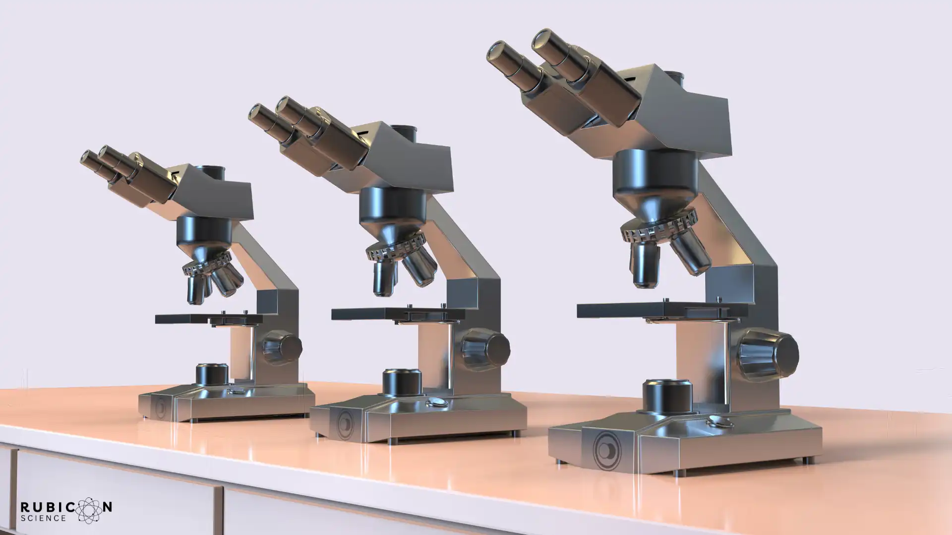
1.1 Optical Microscopes
These classic instruments contain basic lens components to generate 2-dimensional magnified images of transparent specimens, such as fixed cells on glass slides. I always get nostalgic remembering the childhood excitement of peering into school lab microscopes!
Key perks are easy specimen prep, simple operation, and low cost – making optical microscopy ideal for learning environments.
1.2 Stereo or Dissecting Microscopes
Have you tried dissecting tiny insects, plant parts, mechanical objects, or electronics? Stereo microscopes to the rescue! Their defining traits are dual optical paths with separate eyepieces delivering enhanced 3D visualization of solid, opaque samples up to 300x magnification.
The trinocular head design also enables the attachment of external cameras and monitors for sharing observations among groups – perfect for medical and industrial settings.
1.3 Digital Microscopes
Tired of squinting through uncomfortable eyepieces or juggling bulky camera add-ons? Digital microscopes come fully integrated with a display screen, leveraging cutting-edge optics, responsive sensors, and user-friendly software for seamless image capture, measurement, and analysis.
And the benefits don’t end here! Their compact, portable profile suits field applications with challenging lighting or positioning. Fancy some microbe hunting in exotic locales? Digital’s the way to go!
2. Electron Microscopes – Unlocking Atomic Worlds
But optical instruments ultimately hit an impasse. How could researchers possibly examine smaller specimens like viruses, molecules, or crystal defects? The solution came in 1925 via electron microscopes and their Nobel prize-winning development.
Instead of visible light waves, electron microscopes utilize focused beams of electrons and their much shorter wavelengths to visualize submicron-level atomic worlds – with magnifications 100-1,000,000 times greater than light versions! Let’s explore some mainstream electron microscope families a bit further.
2.1 Scanning Electron Microscopes (SEM)
Meet the go-to device for ultra-detailed 3D imaging of cell surfaces, tissues, insects, organs, metals, nanostructures, and more at the micron-to-nanometer scale. SEMs scan samples with a zigzag electron beam, detecting signals that reveal information about topography and composition.
But how does it construct such highly magnified micrographs? Stay with me here… when the incident electrons hit atoms in the specimen, they scatter back distinctive secondary and backscattered electrons. Detectors collect these emitted particles, correlating them to the surface properties of specific scanned regions.
Advanced SEMs can achieve resolution below 1 nm – the breadth of a DNA helix!
2.2 Transmission Electron Microscopes (TEM)
While SEMs visualize exterior sample textures, TEMs shine beams through ultrathin (50-500 nm) sections to intricately map internal microanatomy like cell organelles, proteins, nucleic acids, or nanoparticle innards.
The electrons that transmit contain structural and compositional clues. But another Nobel-worthy innovation – electron diffraction – expanded analytical capabilities even further. Can you guess how crystals were originally discovered to have atomic lattice patterns? It was via TEMs revealing regular diffraction spots based on the scattered electron beam direction.
With cutting-edge TEM devices reaching sub-atomic (picometer) resolution, researchers can effectively scan the fine structure of individual atoms!
3. Scanning Probe Microscopes That Brought The Nanotech Revolution
The 1980s ushered in yet another Nobel-honored microscopy achievement via scanning probe microscopes (SPMs). You’re likely asking yourself – how do these complexly-named systems work?
Instead of light or electron beams, SPMs utilize an exceptionally sharp miniature tip just nanometers wide, rastering over sample surfaces similar to SEMs. This skilled tip intricately senses atomic and nano-level forces, structures, properties, or manipulations.
But the power lies in specialized instrumentation turning subtle tip interactions into comprehensive 3D topographic images or analytical maps. Ready to glimpse some of the trailblazers?
3.1 Scanning Tunneling Microscopes (STMs)
Debuting first, STMs pass an ultrafine conductive tip just angstroms over a sample to enable quantum tunneling of electrons between the surfaces. Why is this tiny gap critical? It ensures positions remain stable enough for tip travel while sensing impossibly slight atomic forces.
Next, monitoring electronics record tunneling current fluctuations, intelligently translating them into extractable data on electron density, atomic structure, conductive properties, and more down to 0.1 nm clarity. Talk about mind-blowing nanotech!
3.2 Atomic Force Microscopes (AFMs)
Rounding out our star SPM lineup, we have AFMs. Their claim to fame? No need for conductive samples, electric current, or even vacuum conditions – broadening nano-imaging possibilities to polymers, glass, ceramics, composites, biomaterials, and delicate molecules.
So how does the AFM tip gather intelligence? Spring-mounted with a laser beam bounce system for ultrasensitivity, the tip directly taps sample surfaces, attracting or repelling atomic forces. Tracking accompanying vertical and lateral deflections maps out intricate morphological details down to fractions of a nanometer!
Don’t forget to check out our in-depth comparative guide on STM and AFM.
4. X-Ray Microscopes
What if I told you microscopes exist to visualize worlds opaque even to electron beams? Enter X-ray microscopy, harnessing high energy light beyond the visible and ultraviolet spectrum.
Similar to their counterparts, X-ray microscopes direct focused beams at specimens, producing fluorescence maps, diffraction patterns, or magnified images detailing buried structures and densities.
But the X-ray advantage? Penetrating centimeters deep into solid objects or living tissue with resolutions ranging from 50-100 nm for scanned images. Researchers leaped at the chance to probe mineral compositions, wiring defects, and metal alloy integrity.
I can’t help imagining what biological marvels might still emerge using fluorescent X-ray nanotomography or psychographic scans! Could we trace in vivo neural pathways, vascular networks, or metabolic organelles? The possibilities seem endless…
The catch so far? Limits to beam intensity and imaging fields of view constrain widespread adoption. Nonetheless, the future looks bright in pioneering labs.
5. Super Resolution Microscopy
If traditional optical microscopes can’t capture single protein or RNA strands clearly due to diffraction issues, how will modern biologists advance molecular knowledge? Enter the 21st-century gift of super-resolution or nano-scale light microscopy – crowned by the Nobel Organization in 2014.
How exactly does it shatter stubborn optical limits? A combination of innovative fluorescent probes, ultra-precise beam manipulation, and computationally-intensive imaging algorithms, stacked together for microscopy magic! Let’s spotlight some techniques paving the wave forward:
5.1 Stimulated Emission Depletion (STED) Microscopy
In a nutshell, STED microscopes use two laser pulses to selectively deactivate fluorescent tags at the sample periphery, exponentially narrowing the central beam width to achieve enhanced resolutions below 10 nm!
5.2 Photo-Activated Localization Microscopy (PALM)
Alternatively, PALM instruments activate individual fluorescent molecules randomly over time. The software tracks thousands of sparse light points, triangulating them into a high-resolution final image. Both STED and PALM proved powerful for single protein tracing!
5.3 Stochastic Optical Reconstruction Microscopy (STORM)
If you ask me, STORM systems synergize the advantages of STED speed with PALM precision. They turn tiny subsets of photoswitchable probes bright or dark stochastically, localizing them with algorithms into a complete structure map to the nanometer scale.
With optical super-resolution microscopes hitting the 1-2 nm clarity milestone already, who knows what cellular revelations await?
Scientists predict a better understanding of chromatin folding, membrane protein distribution, RNA transcription dynamics, and early embryonic development.
Conclusion
And there you have it – the 5 most essential, field-advancing microscopes to emerge in scientific history! We traced the journey from the first optical versions to electron and scanning probe devices unlocking atomic-scale worlds ripe for nanotech innovation. Researchers also won’t be hampered by stubborn optical barriers much longer thanks to recent super-resolution breakthroughs.
Each platform came with specific strengths suiting unique applications across industrial, medical, and academic spheres. Nonetheless, our shared mission remains advancing collective knowledge about the building blocks of life, matter, and the universe itself – one magnified image at a time!
I don’t know about you, but glimpsing these hidden microcosms firsthand seems magical to me. We are extraordinarily fortunate microscopy pioneers persevered over decades perfecting such wonderous windows into worlds otherwise invisible.
Which microscopic realms intrigue you most? Delicate neuronal connections firing memories in our brains? The bustling ecosystem held in one water droplet? Snowflakes in their hexagonal diversity? Endless biochemical activities powering our cells? Feel free to share any marvels spotted under the lens!
For all we uncovered, I suspect microscopes will continue playing instrumental roles across scientific disciplines for ages to come. Miniaturization and automation efforts even aim to bring advanced functionality into laboratories worldwide, classrooms, and our homes for streamlined sample testing and telemedicine.
The future holds breathtaking possibilities – with microscopy leading the charge as always! But I hope you walk away today with a deeper appreciation for these taken-for-granted scientific workhorses so vital for probing life’s hidden mysteries one innovative image at a time!