
The oil immersion hanging drop technique involves placing a drop of liquid sample containing microorganisms on a coverslip, inverting the coverslip placing it on a depression slide, and sealing it with petroleum jelly or any other silicone-based immersion oil. The slide is then viewed under a microscope using an oil immersion objective lens with 100x total magnification.
The hanging drop technique allows for long-term analysis of living microbes in a small droplet while preventing dehydration.
Pre-Requisites for Hanging Drop Technique
Here are the key components and steps involved in sample preparation for the hanging drop technique:
1. Liquid Medium
An appropriate liquid microbial culture medium is selected based on the sample type. Common mediums used include broth, saline, and water.
2. Microbial Sample
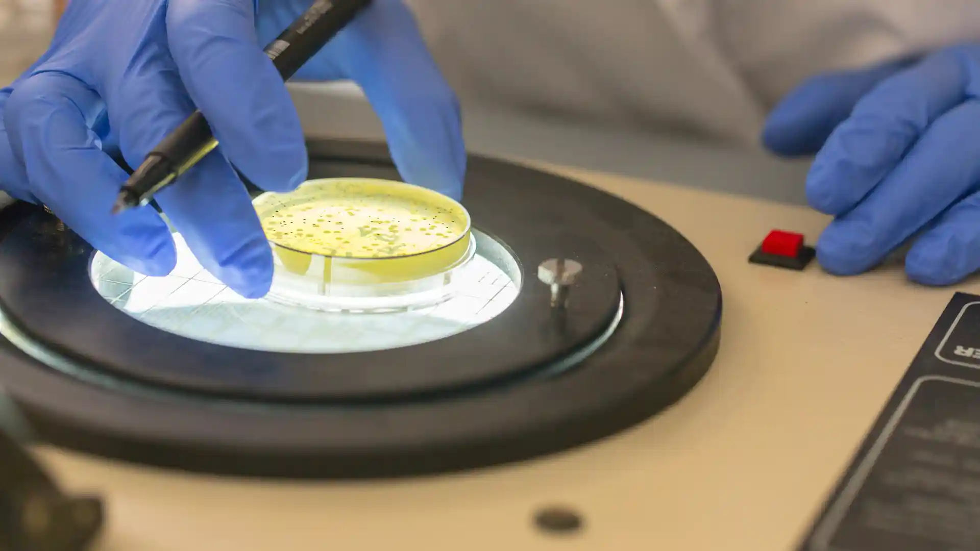
A small sample of the microbial culture or suspension is obtained. Bacterial, yeast, and protozoa cultures are commonly used.
3. Coverslips
Small circular glass coverslips (12-18 mm diameter) are used. Coverslips are cleaned and sterilized before use.
4. Pipettes and tips
Micropipettes and sterile tips are used to handle small liquid volumes. Calibrated pipettes allow precise drop volumes.
5. Sealant
Petroleum jelly or vacuum grease is used to seal the edges of the coverslip to the slide. This prevents sample evaporation.
Step-by-Step Workflow of Hanging Drop Technique

Step 1: Sample Preparation
An appropriate liquid culture medium is selected based on the microbial sample being studied. A small drop of the culture, around 20-50μL, containing the microorganisms is pipetted onto the center of a sterile glass coverslip. The droplet should be large enough to avoid quick evaporation but small enough to confine the microbes.
Step 2: Mounting the Hanging Drop
The coverslip is gently inverted and placed over the concave depression on a glass slide, with the droplet now hanging from the coverslip into the depression. The edges of the coverslip are sealed onto the slide using a thin layer of petroleum jelly like Vaseline. This creates a humid chamber, preventing the sample from drying out.
Step 3: Microscope Setup
The slide is placed on the microscope stage and viewed under a 100x oil immersion objective. Immersion oil is used between the objective lens and the underside of the coverslip to improve resolution. A phase contrast condenser is used to provide good contrast images of transparent microbes.
Applications of the Hanging Drop Technique
The hanging drop technique allows long-term microscopic analysis of living microorganisms, such as:
Microbial Motility
The activity and motility of individual bacterial cells can be observed over time. Flagellar movement and twitching motility can be analyzed.
Microbial Growth
The growth and cell division of microbes can be monitored under controlled conditions. Growth rates and microbial morphology can be studied.
Cellular Secretion
The production and release of metabolic products or enzymes from cells can be observed using suitable stains.
Microbial Interactions
Interactions between multiple microbial species such as symbiosis, predation, and parasitism can be analyzed over time.
Advantages
- Prevents dehydration of sample by creating a humid chamber
- Allows long-term analysis of microbes with good resolution
- Condenser provides high-contrast images
- Small sample size confines microbes for optimal viewing
- Useful for observing microbial motility and growth dynamics
Limitations
- The sample needs to be diluted to an optimal density
- Limited field of view compared to dry mount
- Can only observe one sample at a time
- Risk of contamination while setting up slide
FAQ
How long can microbes be observed using this technique?
With a well-sealed hanging drop, microbes can usually be observed for several hours up to 2-3 days before drying out occurs. Proper sealing is key for long-term analysis.
Does this technique require a special microscope?
A standard compound light microscope with 40x and 100x objective lenses, condenser, and oil immersion functionality is adequate. Phase contrast is optimal but not necessary.